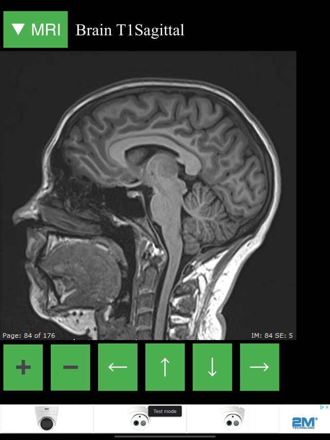
40 brain mri with labels
Arterial Spin Labeling Perfusion of the Brain: Emerging Clinical ... Introduction. Arterial spin labeling (ASL) is a magnetic resonance (MR) imaging technique that enables the measurement of brain perfusion noninvasively at the tissue level.Benefiting from the contrast of inflowing magnetically labeled blood, ASL obviates an exogenous contrast agent. Although the principle of ASL was introduced in early 1990s (1-3) and is feasible on low-field-strength MR ... › pmc › articlesImaging the Addicted Human Brain - PMC - PubMed Central (PMC) Insights From Functional MRI. The differences in brain activity patterns revealed by functional MRI provide invaluable information on a range of issues. Studies have correlated regional brain patterns in response to taking a drug with vulnerability to drug abuse, addictive symptoms and behaviors, and long-term cognitive capacity.
MRI head axial T2 - labeling questions - Radiopaedia The labeled structures are (excluding the correct side): cervical spinal cord posterior arch of C1 odontoid process (peg or dens) of C2 parotid gland intradural segment (V4) of dominant vertebral artery cisterna magna intradural segment (V4) of non-dominant vertebral artery cerebellar tonsil occipital condyle medulla oblongata

Brain mri with labels
MRI head sagittal T1 - labeling questions | Radiology Case ... The labeled structures are (excluding the correct side): temporal horn of lateral ventricle primary fissure of cerebellum choroid plexus trigone (atrium) of lateral ventricle horizontal fissure of cerebellum occipital horn of lateral ventricle intraorbital segment of optic nerve diploic space of parietal bone body of caudate nucleus maxillary sinus 101 Labeled Brain Images and a Consistent Human Cortical Labeling ... Labeling the macroscopic anatomy of the human brain is instrumental in educating biologists and clinicians, visualizing biomedical data, localizing brain data for identification and comparison, and perhaps most importantly, subdividing brain data for analysis. Labeled anatomical subdivisions of the brain enable one to quantify and report brain imaging data within brain regions, which is routinely done for functional, diffusion, and structural magnetic resonance images (f/d/MRI) and positron ... 101 labeled brain images and a consistent human cortical ... - PubMed To manually label the macroscopic anatomy in magnetic resonance images of 101 healthy participants, we created a new cortical labeling protocol that relies on robust anatomical landmarks and minimal manual edits after initialization with automated labels.
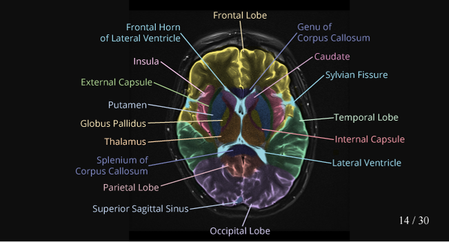
Brain mri with labels. Brain MRI: How to read MRI brain scan | Kenhub Reading time: 20 minutes. Normal brain MRI. A brain MRI is one of the most commonly performed techniques of medical imaging. It enables clinicians to focus on various parts of the brain and examine their anatomy and pathology, using different MRI sequences, such as T1w, T2w, or FLAIR. MRI is used to analyze the anatomy of the brain and to ... Frontiers | 101 Labeled Brain Images and a Consistent Human Cortical ... Labeled anatomical subdivisions of the brain enable one to quantify and report brain imaging data within brain regions, which is routinely done for functional, diffusion, and structural magnetic resonance images (f/d/MRI) and positron emission tomography data. Brain: Atlas of human anatomy with MRI - e-Anatomy - IMAIOS Anatomy of the brain (MRI) - cross-sectional atlas of human anatomy. The module on the anatomy of the brain based on MRI with axial slices was redesigned, having received multiple requests from users for coronal and sagittal slices. The elaboration of this new module, its labeling of more than 524 structures on 379 MRI images in three different views and on 26 anatomical diagrams, took more than 6 months. MRI-labeling: label human brain MRI image by AAL/BA system Input description: x, y, z : x,y,z value of the mni coordinate. distance (default is T): If the MNI coordinate does not belong to any AAL/BA brain region (e.g. white matter, ventricle), then output the closest AAL/BA brain region name and the their distance (mm). When the MNI coordinate does fall into an AAL brain region, then output distance=0.
› user › VideoJugVideojug - YouTube Welcome to Videojug! Here you'll find the best how-to videos around, from delicious, easy-to-follow recipes to beauty and fashion tips. Deep learning to automate the labelling of head MRI datasets for ... In order to generate 'reference-standard image labels' for model testing, 950 head MRI examinations were randomly selected from the 5000 examinations with reference-standard report labels. Two neuroradiologists labelled 250 examinations as normal or abnormal applying the same framework used for report labelling—but interrogating the ... Atlas of BRAIN MRI - W-Radiology The most common MRI sequences used include T1-weighted (T1w) and T2-weighted (T2w) scans (5). T1w sequences display those structures mainly made with fat. Thus, they reveal gray matter as gray, white matter as white, bones as black, and cerebrospinal fluid as black. Meanwhile, T2w sequences highlight structures containing more water. MRI brain (summary) | Radiology Reference Article - Radiopaedia This is a basic article for medical students and other non-radiologists. MRI brain is a specialist investigation that is used for the assessment of a number of neurological conditions. It is the main method to investigate conditions such as multiple sclerosis and headaches, and used to characterize strokes and space-occupying lesions.
Labeled imaging anatomy cases | Radiology Reference Article ... This article lists a series of labeled imaging anatomy cases by body region and modality. Brain CT head: non-contrast axial CT head: non-contrast coronal CT head: non-contrast sagittal CT head: angiogram axial CT head: angiogram coronal CT... Brain lobes - annotated MRI | Radiology Case | Radiopaedia.org Headline: HIV-associated wasting prevalence in the era of modern antiretroviral therapy. Javeeda Siddiqui et al., AIDS, 2022. Favourable outcome of combined medical surgical treatment of cerebral aspergillosis of pulmonary origin. Sergio Ruiz-Santana et al., JMMCR, 2015. MR characteristics of unruptured intracranial arteriovenous malformations ... › createJoin LiveJournal Password requirements: 6 to 30 characters long; ASCII characters only (characters found on a standard US keyboard); must contain at least 4 different symbols; MRI Brain Atlas - University of Minnesota This web app Atlas is intended for veterinary students and radiologists seeking quick access to canine brain anatomy through a mobile device. Via a toggle button, either MRI images or approximately comparable Brain Transection images may be viewed with or without labels. Navigation & Labels.
Labelled MRI of Normal Brain - Stock Image - C017/4421 ...
› instructionsvolBrain: Automated MRI Brain volumetry system 3- lesionBrain pipeline. lesionBrain is a pipeline to automatically segment white matter lesions from MRI data(T1 + FLAIR). As for volBrain, it gets two anonymized MRI brain volumes in NIFTI format and produces a pdf report with the volumes of the lesions and their locations The average processing time of the whole pipeline is around 20 minutes.
fMRI: Arterial Spin Labeling
Automated MRI image labelling processes 100,000 brain exams in under 30 ... Researchers from the School of Biomedical Engineering & Imaging Sciences at King's College London have automated brain MRI image labeling, needed to teach machine learning image recognition...

Brain: Atlas of human anatomy with MRI - e-Anatomy
Head MRI: Purpose, Preparation, and Procedure - Healthline A head MRI is a useful tool for detecting a number of brain conditions, including: aneurysms, or bulging in the blood vessels of the brain; multiple sclerosis; spinal cord injuries; hydrocephalus ...
CaseStacks.com - MRI Brain Anatomy
UCLA Brain Mapping Center - ICBM Template Cortical gyri, subcortical structures and the cerebellum have been delineated from the structural brain template and assigned a unique label. The 3-D set of labels can be imported and registered onto the structural MRI of any individual subject through software like BrainSuite.
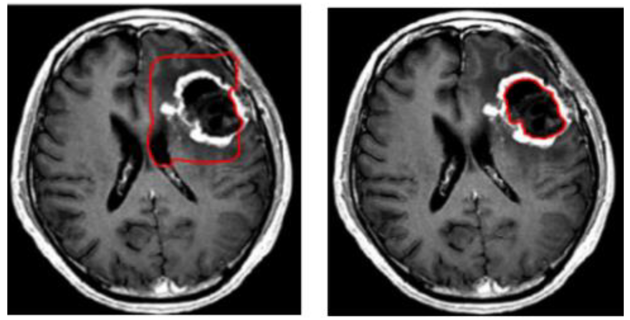
Symmetry | Free Full-Text | 3D-MRI Brain Tumor Detection ...
Labeled MRI Brain Scans - Neuromorphometrics We can also label scans that you provide and we are very interested in labeling white matter anatomy as seen in diffusion-weighted MRI scans. If you want an aggregate version of our data, we can provide it as a probabilistic atlas. The cost to label a single scan is $2449 (USD).

MRI head axial T2 - labeling questions | Radiology Case ...
Brain MRI Images for Brain Tumor Detection | Kaggle Brain MRI Images for Brain Tumor Detection. Brain MRI Images for Brain Tumor Detection. Data. Code (250) Discussion (8) About Dataset. No description available. Health Biology Classification Computer Vision Deep Learning. Edit Tags. close. search. Apply up to 5 tags to help Kaggle users find your dataset.
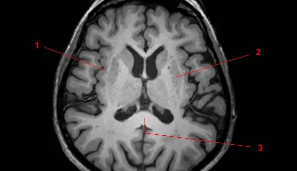
Basal Ganglia Annotated Structures Brain Mri Stock ...
› en › e-AnatomyCross-sectional anatomy of the brain - e-Anatomy - IMAIOS Apr 15, 2022 · Axial MRI Atlas of the Brain. Free online atlas with a comprehensive series of T1, contrast-enhanced T1, T2, T2*, FLAIR, Diffusion -weighted axial images from a normal humain brain. Scroll through the images with detailed labeling using our interactive interface. Perfect for clinicians, radiologists and residents reading brain MRI studies.
T1-weighted in vivo human whole brain MRI dataset with an ...
brain anatomy | MRI coronal brain anatomy | free MRI cross sectional ... ELBOW AXIAL. WRIST AXIAL. WRIST CORONAL. KNEE CORONAL. KNEE SAGITTAL. ARTERIES UPPER LEG. ARTERIES LOWER LEG. This MRI brain coronal cross sectional anatomy tool is absolutely free to use. Use the mouse scroll wheel to move the images up and down alternatively use the tiny arrows (>>) on both side of the image to move the images.
Brain Lesion Detection in MRI Images with Graph-cut Algorithms
MRI Brain Animated Quiz - University of Minnesota MRI Brain Animated Quiz. Canine Brain MRI Anatomy Quiz. Sequentially click/tap: first the dot associated with a term; then, its corresponding target dot on the MRI image. If a line connection appears, your choice was correct! White Matter. Cerebral Cortex. Olfactory Bulb. Longitudinal Fissure.

volBrain: Automated MRI Brain volumetry system
Brain Tumor MRI Classification | VGG16 | Kaggle Brain Tumor MRI Classification | VGG16. Notebook. Data. Logs. Comments (16) Run. 7416.0s - GPU P100. history Version 9 of 9. Cell link copied. License. This Notebook has been released under the Apache 2.0 open source license. Continue exploring. Data. 2 input and 4 output. arrow_right_alt. Logs. 7416.0 second run - successful.

Brain Anatomy MRI- Neuroradiology
Split-Attention U-Net: A Fully Convolutional Network for Robust Multi ... Multi-label brain segmentation from brain magnetic resonance imaging (MRI) provides valuable structural information for most neurological analyses. Due to the complexity of the brain segmentation algorithm, it could delay the delivery of neuroimaging findings. Therefore, we introduce Split-Attention …

Cross-sectional anatomy of the brain - e-Anatomy
Brain MRI: What It Is, Purpose, Procedure & Results - Cleveland Clinic A brain MRI (magnetic resonance imaging) scan, also called a head MRI, is a painless procedure that produces very clear images of the structures inside of your head — mainly, your brain. MRI uses a large magnet, radio waves and a computer to produce these detailed images. It doesn't use radiation. Currently, MRI is the most sensitive imaging test of your head (particularly, your brain), as compared to other imaging techniques, such as CT (computed tomography) scans or X-rays.

Automated segmentation of the hypothalamus and associated ...
MRI anatomy | free MRI axial brain anatomy - Mrimaster.com WRIST AXIAL. WRIST CORONAL. KNEE CORONAL. KNEE SAGITTAL. ARTERIES UPPER LEG. ARTERIES LOWER LEG. This MRI brain cross sectional anatomy tool is absolutely free to use. Use the mouse scroll wheel to move the images up and down alternatively use the tiny arrows (>>) on both side of the image to move the images.

MRI anatomy | free MRI axial brain anatomy
en.wikipedia.org › wiki › NeuroimagingNeuroimaging - Wikipedia Computed tomography (CT) or Computed Axial Tomography (CAT) scanning uses a series of x-rays of the head taken from many different directions. Typically used for quickly viewing brain injuries, CT scanning uses a computer program that performs a numerical integral calculation (the inverse Radon transform) on the measured x-ray series to estimate how much of an x-ray beam is absorbed in a small ...
Recommended implementation of arterial spinв•'labeled ...
› articles › s41598/021/90428-8Brain tumor segmentation based on deep learning and an ... May 25, 2021 · Brain tumor localization and segmentation from magnetic resonance imaging (MRI) are hard and important tasks for several applications in the field of medical analysis. As each brain imaging ...

CerebrA: Accurate registration and manual label correction of ...
Brain MRI segmentation | Kaggle Journal of Neuro-Oncology, 2017. This dataset contains brain MR images together with manual FLAIR abnormality segmentation masks. The images were obtained from The Cancer Imaging Archive (TCIA). They correspond to 110 patients included in The Cancer Genome Atlas (TCGA) lower-grade glioma collection with at least fluid-attenuated inversion ...
Magnetic resonance imaging of the brain - Wikipedia
Manually Labeled MRI Brain Scan Database - nitrc.org Image 1 of 3. Click for more. This is a continuously growing and improving database of high-quality neuroanatomically labeled MRI brain scans, created not by an algorithm, but by neuroanatomical experts. All results are checked and corrected. Regions of interest include the usual sub-cortical structures (thalamus, caudate, putamen, hippocampus ...

How much does a brain MRI cost? | From $225
101 labeled brain images and a consistent human cortical ... - PubMed To manually label the macroscopic anatomy in magnetic resonance images of 101 healthy participants, we created a new cortical labeling protocol that relies on robust anatomical landmarks and minimal manual edits after initialization with automated labels.

Cross-sectional anatomy of the brain - e-Anatomy
101 Labeled Brain Images and a Consistent Human Cortical Labeling ... Labeling the macroscopic anatomy of the human brain is instrumental in educating biologists and clinicians, visualizing biomedical data, localizing brain data for identification and comparison, and perhaps most importantly, subdividing brain data for analysis. Labeled anatomical subdivisions of the brain enable one to quantify and report brain imaging data within brain regions, which is routinely done for functional, diffusion, and structural magnetic resonance images (f/d/MRI) and positron ...

Region Of Interest Based Image Classification: A Study in MRI ...
MRI head sagittal T1 - labeling questions | Radiology Case ... The labeled structures are (excluding the correct side): temporal horn of lateral ventricle primary fissure of cerebellum choroid plexus trigone (atrium) of lateral ventricle horizontal fissure of cerebellum occipital horn of lateral ventricle intraorbital segment of optic nerve diploic space of parietal bone body of caudate nucleus maxillary sinus
MRI of the White and Gray Matter in the Brain - W-Radiology
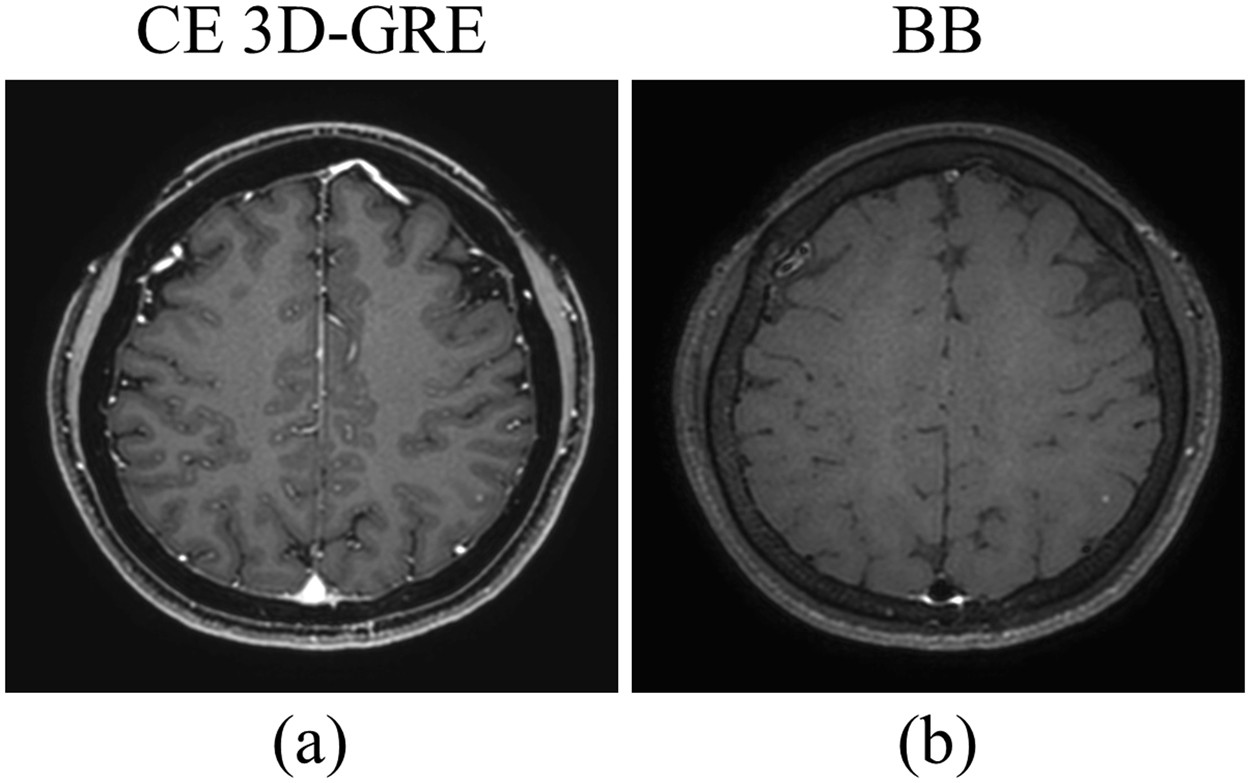
Deep-learned 3D black-blood imaging using automatic labelling ...
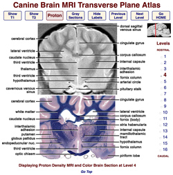
Veterinary Planar Anatomy Courseware
Magnetic resonance image (MRI) of a side view of the brain ...
Review of “Brain MRI Atlas” App for the iPad | SpringerLink

volBrain: Automated MRI Brain volumetry system

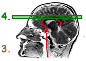
Read on for my tips at looking at a sagittal view MRI of the ...

How To Read A Brain MRI Radiology Report - Part II | Blog ...

Label Each Part of the Brain Scan | MS in African Americans ...

MRI anatomy | free MRI axial brain anatomy
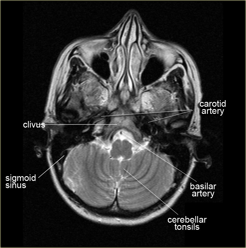
The Radiology Assistant : Anatomy
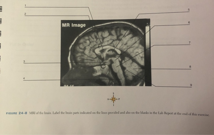
Solved MR Image Up FIGURE 24-8 MRI of the brain. Label the ...

Brain Anatomy and Images Brain

Cross sectional Anatomy of Brain on... - World Of Radiology ...

Brain Tumor Detection and Localization - Analytics Vidhya

MRI anatomy | free MRI axial brain anatomy

7.0 T MRI Axial Images. (a) An axial view image obtained by ...

MRI Scans Show The Horrific Effect Cocaine Abuse Can Have On ...

Anatomy of the brain: T2 weighted magnetic resonance image ...
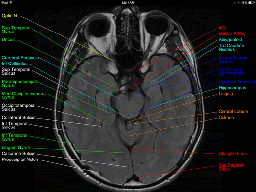
MRI Viewer on the App Store
Post a Comment for "40 brain mri with labels"