38 microscope diagram without labels
Fluorescence - Wikipedia Fluorescence is the emission of light by a substance that has absorbed light or other electromagnetic radiation.It is a form of luminescence.In most cases, the emitted light has a longer wavelength, and therefore a lower photon energy, than the absorbed radiation.A perceptible example of fluorescence occurs when the absorbed radiation is in the ultraviolet region of the … Preface | Python Data Science Handbook - GitHub Pages Mar 26, 2013 · While some of the intersection labels are a bit tongue-in-cheek, this diagram captures the essence of what I think people mean when they say "data science": it is fundamentally an interdisciplinary subject. Data science comprises three distinct and overlapping areas: the skills of a statistician who knows how to model and summarize datasets (which are …
Microorganisms: Friend and Foe Class 8 Extra Questions Oct 11, 2019 · Pull out a gram or bean plant from the field. Observe its roots. You will find round struc¬tures called root nodules on the roots. Draw a diagram of the root and show the root nod¬ules. Answer: Question 2. Collect the labels from the bottles of jams and jellie on the labels. Answer: Do it yourself. Question 3. Visit a dcotor.

Microscope diagram without labels
Fluorescence In Situ Hybridization (FISH) | Learn Science at Scitable Cytogenetics entered the molecular era with the introduction of in situ hybridization, a procedure that allows researchers to locate the positions of specific DNA … Fluorescence Resonance Energy Transfer (FRET) Microscopy Presented in Figure 3 is a Jablonski diagram illustrating the coupled transitions involved between the donor emission and acceptor absorbance in fluorescence resonance energy transfer. Absorption and emission transitions are represented by straight vertical arrows (green and red, respectively), while vibrational relaxation is indicated by wavy ... Multiphoton Microscopy | Nikon’s MicroscopyU Two-photon excitation microscopy (also referred to as non-linear, multiphoton, or two-photon laser scanning microscopy) is an alternative to confocal and deconvolution microscopy that provides distinct advantages for three-dimensional imaging.In particular, two-photon excitation excels at imaging of living cells, especially within intact tissues such as brain slices, embryos, …
Microscope diagram without labels. Welcome to Butler County Recorders Office Copy and paste this code into your website. Your Link Name Looking at the Structure of Cells in the Microscope A typical animal cell is 10–20 μm in diameter, which is about one-fifth the size of the smallest particle visible to the naked eye. It was not until good light microscopes became available in the early part of the nineteenth century that all plant and animal tissues were discovered to be aggregates of individual cells. This discovery, proposed as the cell doctrine by Schleiden and … Electron microscope - Wikipedia An electron microscope is a microscope that uses a beam of accelerated electrons as a source of illumination. As the wavelength of an electron can be up to 100,000 times shorter than that of visible light photons, electron microscopes have a higher resolving power than light microscopes and can reveal the structure of smaller objects.. Electron microscopes use shaped magnetic … mastering ch.13 Flashcards & Practice Test | Quizlet Complete the diagram to show the life cycle of a typical animal. Follow these steps: 1. First, drag blue labels onto blue targets only to identify each stage of the life cycle. 2. Next, drag pink labels onto pink targets only to identify the process by which each stage occurs. 3.
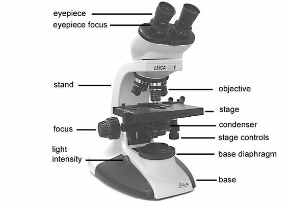
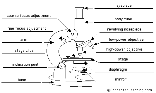
Compound Microscope Parts, Functions, and Labeled Diagram Nov 18, 2020 · Common compound microscope parts include: Compound Microscope Definitions for Labels Eyepiece (ocular lens) with or without Pointer: The part that is looked through at the top of the compound microscope. Eyepieces typically have a magnification between 5x & 30x. Microscope Objective Lens | Products | Leica Microsystems The objective lens is a critical part of the microscope optics. The microscope objective is positioned near the sample, specimen, or object being observed. It has a very important role in imaging, as it forms the first magnified image of the sample. The numerical aperture (NA) of the objective indicates its ability to gather light and largely determines the microscope’s resolution, the ... Dijkstra's algorithm - Wikipedia Dijkstra's algorithm (/ ˈ d aɪ k s t r ə z / DYKE-strəz) is an algorithm for finding the shortest paths between nodes in a graph, which may represent, for example, road networks.It was conceived by computer scientist Edsger W. Dijkstra in 1956 and published three years later. Wikipedia:Citation needed - Wikipedia A complete version of the documentation for this template is provided at Template:Citation needed.If you are new to editing and instead just need a general overview of how sources work, please visit the referencing for beginners help page.
CODEX multiplexed tissue imaging with DNA-conjugated antibodies - Nature Jul 02, 2021 · This protocol describes co-detection by indexing, a highly multiplexed imaging technology that uses DNA-conjugated antibodies to image up to 60 markers in formalin-fixed, paraffin-embedded and ... Multiphoton Microscopy | Nikon’s MicroscopyU Two-photon excitation microscopy (also referred to as non-linear, multiphoton, or two-photon laser scanning microscopy) is an alternative to confocal and deconvolution microscopy that provides distinct advantages for three-dimensional imaging.In particular, two-photon excitation excels at imaging of living cells, especially within intact tissues such as brain slices, embryos, … Fluorescence Resonance Energy Transfer (FRET) Microscopy Presented in Figure 3 is a Jablonski diagram illustrating the coupled transitions involved between the donor emission and acceptor absorbance in fluorescence resonance energy transfer. Absorption and emission transitions are represented by straight vertical arrows (green and red, respectively), while vibrational relaxation is indicated by wavy ... Fluorescence In Situ Hybridization (FISH) | Learn Science at Scitable Cytogenetics entered the molecular era with the introduction of in situ hybridization, a procedure that allows researchers to locate the positions of specific DNA …
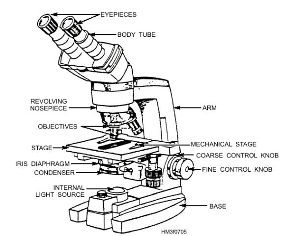
Compound Microscope Diagram With Labels - Micropedia
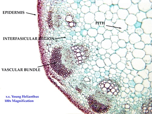
Biology 252 -- Plant Morphology and Systematics Home Page
8 Best Images of Lens Diagram Worksheet - Microscope with Labeled Parts, Label Eye Parts ...

Simple Microscope Labeled Diagram - Micropedia
8 Best Images of Lens Diagram Worksheet - Microscope with Labeled Parts, Label Eye Parts ...
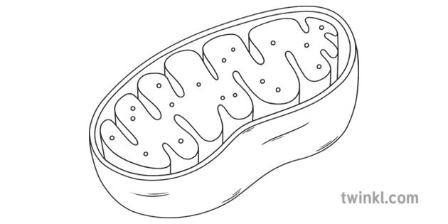
Mitochondria Science Cell Diagram Beyond Black and White RGB Illustration
The Microscope - General Revision for GCSE

Simple Unlabelled Microscope Diagram - Micropedia

Best 110 Histology - Skin images on Pinterest | Anatomy, Anatomy reference and Hair follicles

33 Microscope Diagram To Label - Labels Database 2020
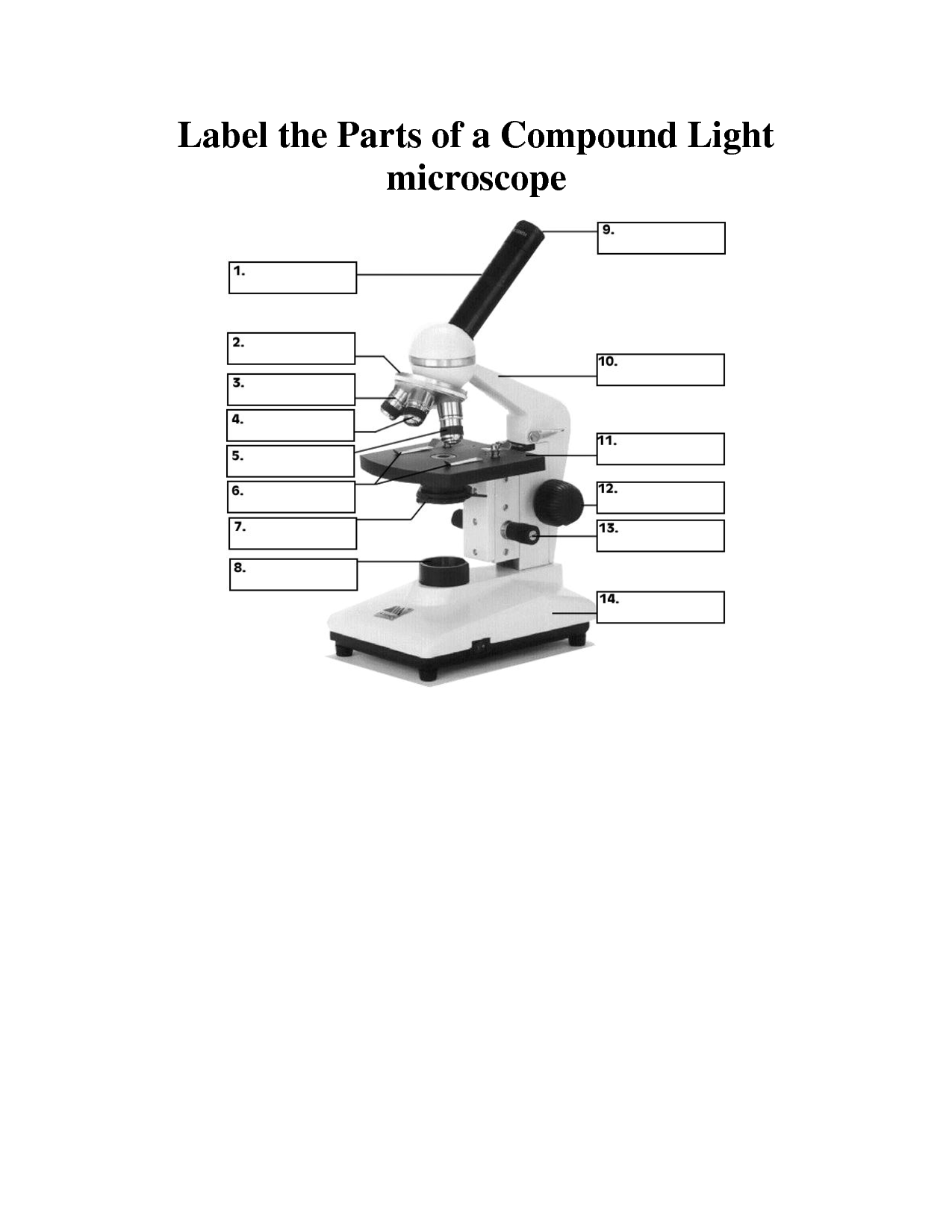
[Solved] 1. Label the following microscope using the components described within the ...
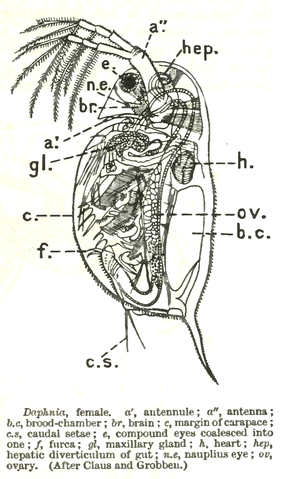
Water flea - Experiments on Microscopes 4 Schools

Human Bio: February 2008

Microscope With Labels Clip Art at Clker.com - vector clip art online, royalty free & public domain

Label diagram of compound microscope - Science - The Fundamental Unit of Life - 12499729 ...

Microscope Unlabeled Diagram - Micropedia

Easy Microscope Labeled Diagram - Micropedia
Post a Comment for "38 microscope diagram without labels"